What is hypoxemia
Hypoxemia is a pathological condition characterized by a decrease in the oxygen content in arterial blood.
Blood passing through the pulmonary vessels is enriched with oxygen and gets rid of carbon dioxide
This process occurs due to the fact that oxygen, for some reason, cannot enter the bloodstream.
Hypoxemia is often confused with hypercapnia (increased partial pressure of carbon dioxide), as well as hypoxia (insufficient supply of oxygen to tissues), although all these are aspects of one general process. Hypoxemia is one of the most common causes of hypoxia.
A holy place is never empty
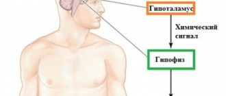
Hypercapnia is an increase in the level of carbon dioxide (CO2) in the blood, hypoxemia is a decrease in the oxygen content (O2) in the same place. How and why does this happen?
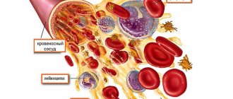
It is known that the transport of oxygen from the lungs with arterial blood is carried out by red blood cells (erythrocytes), where oxygen is bound (but not very tightly) to chromoprotein (hemoglobin). Hemoglobin (Hb), which carries oxygen to the tissues (oxyhemoglobin), upon arrival at its destination, gives up O2 and becomes reduced hemoglobin (deoxyhemoglobin), capable of attaching the same oxygen, carbon dioxide, and water. But since carbon dioxide is already waiting for it in the tissues, which needs to be delivered with venous blood to the lungs for removal from the body, hemoglobin takes it away, turning into carbohemoglobin (HbCO2) - also a fragile compound. Carbohemoglobin in the lungs will break down into Hb, which can combine with oxygen received during inhalation, and carbon dioxide, intended for removal from the body during exhalation.
These reactions can be schematically represented in the form of chemical reactions, which the reader may well remember from school lessons:
- Hb (in red blood cells) + O2 (comes with inhalation with air) → HbO2 – the reaction occurs in the lungs, the resulting compound is sent to the tissues;
- HbO2 → Hb (deoxyhemoglobin) + O2 – in tissues that receive oxygen for respiration;
- Hb + CO2 (waste, from tissues) → HbCO2 (carbohemoglobin) – in the tissues, the formed carbohemoglobin is sent to the small circle for gas exchange and oxygen enrichment;
- HbCO2 (from tissues) → to the lungs: Hb (free to receive oxygen) + CO2↑ (removed with exhalation);
- Hb + O2 (from inhaled air) – new cycle.
However, it should be noted that everything works out so well when there is enough oxygen, there is no excess carbon dioxide, everything is fine with the lungs - the body breathes clean air, the tissues receive everything they should, they do not experience oxygen starvation, the CO2 formed during gas exchange safely leaves organism. The diagram shows that reduced hemoglobin (Hb), without having strong bonds, is always ready to attach any of the components (whatever comes across, it attaches). If at that moment there is less oxygen in the lungs than hemoglobin can take (hypoxemia), and there is more than enough carbon dioxide (hypercapnia), then it will take it (CO2) and carry it to the tissues with arterial blood (arterial hypoxemia) instead of the expected oxygen. Reduced tissue oxygenation is a direct path to the development of hypoxia, that is, oxygen starvation of tissues.
Obviously, it is difficult to separate symptoms such as hypoxia, hypercapnia and hypoxemia - they underlie the development of acute respiratory failure and determine the clinical picture of ARF.
Causes and mechanism of occurrence
Since hypoxemia is a pathological symptom that most often leads to such a functional disorder as hypoxia, the main mechanisms for the development of a decrease in the partial pressure of oxygen in the blood should be noted.
Among the large number of causes and processes, 4 basic development mechanisms can be distinguished:
- A decrease in the partial pressure of oxygen in the environment and, as a result, in the person who is in it. This leads to insufficient oxygenation of the blood. This can happen due to a fire, due to a gas leak, during a long stay in a small space with poor ventilation, or when climbing mountains.
- Hypoventilation (insufficient respiratory movements of the lungs due to depression of the respiratory center, airway obstruction, weakness of nerves and muscles).
- Dissonance between the processes of perfusion and the ventilation mechanism in the lungs. Occurs against the background of decreased ventilation under the condition of normal blood flow or vice versa (oncopathology, pneumonia, a group of chronic obstructive pulmonary diseases).
- Atypical blood shunt from right to left. In patients (usually children) who have congenital heart defects, a pathological discharge of venous blood from the right side of the heart occurs directly into the aorta, bypassing the lungs, where the blood must be saturated with oxygen.
Types of hypoxia developing as a result of hypoxemia, by time of occurrence and duration of course
Factors inhibiting oxygenation
The basis of hypoxemia is a disorder of oxygen saturation of arterial blood in the lungs. You can find out that the blood in the lungs is not oxygenated by such an indicator as the partial oxygen tension (PaO2), the values of which normally should not fall below 80 mm. rt. Art.
The reasons for decreased blood oxygenation are:
- Alveolar hypoventilation, which occurs as a result of the influence of various factors, primarily, a lack of oxygen in the inhaled air, which leads to its decrease in the alveoli and leads to the development of exogenous hypoxia;
- Disorders of ventilation-perfusion ratios that occur in chronic lung diseases are the most common causative factor in the development of hypoxemia and respiratory hypoxia;
- Shunting from right to left in case of circulatory disorders and venous blood entering directly into the left heart without visiting the lungs (heart defects) with the development of circulatory hypoxia;
- Violation of the diffusion abilities of the alveolar-capillary membrane.
So that the reader can imagine the role of the ventilation-perfusion relationship and the importance of the diffusion abilities of the alveolar-capillary membrane, the essence of these concepts should be clarified.
What happens in the lungs?
In the human lungs, gas exchange is ensured by ventilation and blood flow in a small circle, but ventilation and perfusion do not occur to the same extent. For example, certain zones are ventilated, but not provided with blood, that is, they do not participate in gas exchange or, conversely, in some areas the blood flow is preserved, but they are not ventilated and are also excluded from the gas exchange process (alveoli of the apex of the lungs). The expansion of zones not involved in gas exchange (lack of perfusion) leads to hypoxemia, which a little later will lead to hypercapnia.
Impairment of pulmonary blood flow results from various pathological conditions of vital organs and, first of all, the circulatory system, which become causes of hypoxemia:
An example of the development of hypoxemia during pulmonary embolismPulmonary hypertension;
- Acute blood loss;
- Dehydration;
- State of shock of various origins;
- DIC syndrome with the formation of microthrombi in the bloodstream;
- TELA (small branches);
- Pathological conditions with damage to the walls of the pulmonary vessels (vasculitis).
The diffusion capacity of the alveolar-capillary membrane, depending on many parameters, can change its values (increase and decrease) depending on the circumstances (compensatory and adaptive mechanisms during load, changes in body position, etc.). In young adults (over 20 years), it decreases naturally, which is considered a physiological process. An excessive decrease in this indicator is observed in diseases of the respiratory system (pneumonia, edema, COPD, emphysema), which significantly reduce the diffusion capacity of ACM (gases cannot overcome long paths formed as a result of pathological changes, and blood flow is impaired due to a decrease in the number of capillaries) . Due to such disturbances, the main signs of hypoxia, hypoxemia and hypercapnia begin to appear, indicating the development of respiratory failure.
Signs of decreased O2 in the blood
Signs of a decrease in oxygen can appear quickly (oxygen concentration drops, but the body tries to compensate for the loss on its own) or delay (against the background of chronic pathology of the main life support systems, the compensatory capabilities of which have already ended).
Symptoms of hypoxemia:
- Blueness of the skin (cyanosis). The color of the skin determines the severity of the condition, therefore, with a weak degree of hypoxemia, it usually does not reach cyanosis, but pallor, nevertheless, occurs;
- Rapid heartbeat (tachycardia) – the heart tries to compensate for the lack of oxygen;
- Decreased blood pressure (hypotension);
- Fainting if PaO2 drops to very low values (less than 30 mmHg)
A decrease in oxygen concentration in the blood, of course, leads to brain suffering with memory impairment, weakened concentration, sleep disorders (night apnea and its consequences), and the development of chronic fatigue syndrome.
Symptoms and signs
Hypoxemia is an insidious condition that manifests itself only after some time. Symptoms can be divided into several groups. At the very beginning, the body tries to adapt to the lack of oxygen; early signs will allow you to suspect hypoxemia:
- rapid heartbeat (tachycardia);
- pale skin;
- increased blood pressure.
Mechanisms in the body that ensure normal functioning over a period of time and cause characteristic manifestations
All these are manifestations of a compensatory mechanism aimed at maintaining a sufficient amount of oxygen. This allows the body to function as before, but if the cause of hypoxia is not eliminated, hypoxemia will begin to progress. Late symptoms:
- blue discoloration of the skin (cyanosis) first around the lips, on the earlobes, on the tips of the fingers, then throughout the body;
- cold clammy sweat;
- dizziness (up to loss of consciousness);
- euphoria and motor restlessness, followed by stupor;
- neurological disorders.
Features of the course of hypoxemia in pregnant women
When a woman is preparing to become a mother, many functional processes occur in her body that allow her to bear and give birth to a healthy baby. The fetus receives blood enriched with oxygen through the placenta, so if a woman experiences hypoxemia, this can cause pathological (often fatal) complications such as fetal hypoxia and fetoplacental insufficiency. The latter, in turn, can lead to placental abruption and miscarriage.
There are acute and chronic forms of fetal hypoxia (as a consequence of hypoxemia).
Diseases of the expectant mother, which, as a rule, provoke a dangerous condition, include:
- anemia;
- pathologies of the cardiovascular system (arterial hypertension, heart disease, preeclampsia, eclampsia);
- chronic kidney disease;
- diabetes;
- smoking (including passive smoking);
- diseases of the bronchopulmonary system;
- pathology of the placenta and entanglement of the fetus with the umbilical cord;
- post-term pregnancy;
- premature labor.
Symptoms in pregnant women are similar to the manifestations of hypoxemia in adults, but the first thing the expectant mother will pay attention to is the pathological behavior of the fetus (motor movements become less, the woman experiences discomfort in the lower abdomen).
In this case, hospitalization is indicated. Already during the ultrasound procedure, the condition of the child will be visible. Before this, the obstetrician-gynecologist will listen to the fetal heart rate, which is reduced during hypoxia. According to the doctor’s decision, delivery is carried out urgently, because in this condition the baby can die quite quickly.
Fetal hypoxia - video
Important aspects of the manifestation of hypoxemia in children
Hypoxemia in a newborn baby is accompanied by the following signs:
- irregular breathing with periods of its absence (apnea);
The pause in breathing can be 15–20 seconds.
- bluish (cyanotic) skin tone;
- inhibition of motor activity and sucking reflex.
Such a violation requires immediate help, as it can lead to oxygen starvation of the brain and sudden cardiac death. If we talk about a chronic process, then the child will have all the manifestations of intrauterine growth retardation.
If this process is not associated with the pathology of pregnancy, but was developed independently in the child, then you should think about congenital heart defects, which most often manifest themselves as persistent hypoxemia. At the same time, the child becomes restless and aggressive, which after a while can give way to apathy and stupor. In addition, shortness of breath and cyanotic (blue) skin are observed.
With some congenital heart defects, the child experiences so-called dyspnea-cyanotic attacks, during which he squats and breathes heavily. This body position allows the baby to feel easier by increasing the outflow of blood from the veins of the legs.
Acute heart failure in children
Depending on the type of hemodynamics and some features of pathogenesis, the following clinical variants of acute heart failure (AHF) are distinguished:
- with a stagnant type of hemodynamics: right ventricular (venous stagnation in the systemic circulation);
- left ventricular (cardiac asthma, pulmonary edema);
CARDIOGENIC SHOCK (low cardiac output syndrome)
Shock is an acutely developing, life-threatening pathological process characterized by a progressive decrease in tissue perfusion, severe disturbances in the functioning of the central nervous system, blood circulation, respiration and metabolism.
Cardiogenic shock is a clinical syndrome characterized by arterial hypotension and signs of a sharp deterioration in microcirculation and tissue perfusion, including blood supply to the brain and kidneys (lethargy or agitation, drop in diuresis, cold skin covered with sticky sweat, pallor, marbled skin pattern); sinus tachycardia is compensatory in nature.
In addition to cardiogenic shock, possible causes of shock may include:
- A decrease in total blood volume (hypovolemic shock) due to bleeding or dehydration due to losses from the gastrointestinal tract (vomiting, diarrhea), polyuria, burns, etc. The main pathogenetic mechanism is insufficient cardiac preload due to a deficiency of venous inflow.
- Deposition of blood in the venous pools (distributive or vasogenic shock) during anaphylaxis, acute adrenal insufficiency, sepsis, neurogenic or toxic shock. The leading pathogenetic mechanism is insufficiency of cardiac afterload.
Low cardiac output (cardiogenic shock) develops as a result of failure of the pumping function of the heart (acute myocardial ischemia, infectious and toxic carditis, cardiomyopathies), as well as as a result of obstruction of the venous inflow to the heart or cardiac output (obstructive shock) in diseases of the pericardium (pericardial tamponade), tension pneumothorax, acute obstruction of the atrioventricular orifice by atrial myxoma, rupture of chords, heart valves, massive pulmonary embolism, etc. Pericardial tamponade and obstruction of the atrioventricular orifice require immediate surgical intervention; drug therapy in these cases can only worsen the situation.
A special clinical variant of cardiogenic shock is arrhythmic shock, which develops as a result of a drop in cardiac output due to tachycardia/tachyarrhythmia or bradycardia/bradyarrhythmia; After stopping the rhythm disturbance, adequate hemodynamics are quickly restored.
With the development of low cardiac output syndrome, a pain syndrome is observed, manifested by severe anxiety in the child, followed by lethargy. A drop in blood pressure, threadlike pulse, tachycardia, “marble” pallor of the skin, collapsed peripheral veins, sticky cold sweat, acrocyanosis, and oligoanuria are noted.
The course of cardiogenic shock is often accompanied by the development of pulmonary edema, mesenteric ischemia, disseminated intravascular coagulation syndrome, and renal failure.
Cardiogenic shock at the prehospital stage is diagnosed based on:
- progressive drop in systolic blood pressure;
- decrease in pulse pressure - less than 20 mm Hg. Art.;
- signs of impaired microcirculation and tissue perfusion - a drop in urine output of less than 20 ml/h, cold skin covered with sticky sweat, pallor, marbled skin pattern, in some cases - collapsed peripheral veins.
Treatment of cardiogenic shock
- Elimination of the main cause: relief of heart rhythm disturbances and pain. In case of severe pain, administer fentanyl at a dose of 0.01 mg/kg or 1% promedol at a dose of 0.1 ml/year of life intravenously. For children in the first two years of life, prescribe non-narcotic analgesics: baralgin or a 50% analgin solution at a dose of 0.1-0.2 ml/year of life. In the presence of psychomotor agitation, prescribe a 0.5% solution of diazepam (Seduxen, Relanium) at a dose of 0.1-0.3 mg/kg intravenously.
- In the absence of signs of congestive heart failure (shortness of breath, moist rales in the posterior lower parts of the lungs), the patient must be placed in a horizontal position.
- If the clinical picture of shock is extensive and there are no signs of congestive heart failure, therapy should begin with intravenous fluid administration (infusion therapy to increase preload) under the control of blood pressure, heart rate, respiratory rate and auscultation of the lungs. Rheopolyglucin is administered at a dose of 5-8 ml/kg + 10% glucose solution and 0.9% sodium chloride solution at a dose of 50 ml/kg in a ratio of 2 to 1 with the addition of cocarboxylase and 7.5% potassium chloride solution at a dose of 2 mmol/ kg body weight.
- An increase in cardiac output is achieved:
- administration of dopamine (6-9 mg/kg/min), which has a positive inotropic effect. Dopamine, an agonist of dopamine receptors, causes excitation of α- and β-adrenergic receptors, increases the release of norepinephrine into the synaptic cleft, increases the strength of heart contractions and cardiac output, the effect of the drug on heart rate is insignificant. The drug promotes the redistribution of total vascular peripheral resistance, causing dilatation of the renal and mesenteric vessels and a vasoconstrictor effect; improved renal perfusion helps to increase diuresis. Dopamine infusion is carried out in the intensive care unit under continuous monitoring using a dispenser for 24-48 hours. The effect occurs after 5 minutes, its peak after 5-7 minutes. Considering the possible tachycardic and arrhythmogenic effect of dopamine, the drug is used in very short courses, only in extremely severe cases and with complete depletion of the sympathetic-adrenal system, with an increase in AHF to degree III.
- administration of drugs with a positive chronotropic effect: adrenaline, norepinephrine (0.05-0.2 mcg/kg/min).
- The lack of effect from dopamine or the impossibility of its use due to tachycardia, arrhythmia is an indication for the addition or monotherapy with dobutamine, which, unlike dopamine, has a more pronounced vasodilatory effect and a less pronounced ability to cause an increase in heart rate and arrhythmia.
250 mg of the drug is diluted in 500 ml of a 5% glucose solution (1 ml of the mixture contains 0.5 mg, and 1 drop - 25 mcg of dobutamine); in monotherapy, it is prescribed at a dose of 2.5 mcg/kg/min, increasing every 15-30 minutes by 2.5 mcg/kg/min until an effect, side effect is obtained, or a dose of 10 mcg/kg/min is achieved, and with a combination of dobutamine with dopamine - in maximum tolerated doses. Dobutamine is a β1-adrenergic agonist, has a positive inotropic effect on the heart, moderately increases heart rate, as well as stroke and cardiac output, reduces the total peripheral and vascular resistance of the pulmonary circulation, while systemic blood pressure tends to increase, reduces the filling pressure of the ventricles of the heart, increases coronary blood flow, improves oxygen supply to the myocardium. Increased cardiac output improves renal perfusion and increases sodium and water excretion. The drug is used for reduced renal blood flow and cardiac output, moderate hypotension. Dobutamine is not prescribed for systolic blood pressure <70 mm Hg. Art. - If an increase in the volume of administered fluid does not lead to an increase in cardiac output (with high central venous pressure and no effect of drugs with a positive inotropic effect), the reason for its decrease may be either a large afterload, to reduce which sodium nitroprusside is used (0.5-0. 8 mcg/kg/min); or a decrease in myocardial contractility, which is an indication for the administration of cardiac glycosides.
- If there are signs of congestive heart failure and in the case of using inotropic drugs from the group of pressor amines, the administration of peripheral vasodilators - nitrates (nitroglycerin) is indicated.
- Administer a 3% solution of prednisolone at a dose of 3-5 mg/kg intravenously.
- In the absence of contraindications, heparin is prescribed to correct microcirculatory disorders, especially with long-term intractable shock.
- Oxygen therapy.
- Admission to the intensive care unit.
ACUTE CONGESTIVE HEART FAILURE
There are 3 stages of development of acute congestive heart failure.
Stage I:
- decrease in cardiac output, hypervolemia, signs of fluid stagnation in one or both circulations.
- tachycardia and shortness of breath at rest, not consistent with fever,
- change in the ratio between h.s.s. and ch.d. (in children of the first year of life - more than 3.5:1; older than one year - more than 4.5:1),
- increase in liver size,
- periorbital edema (mainly in the first year),
- swelling of the neck veins and puffiness of the face,
- cyanosis,
- bronchospasm,
- silent crepitating or fine rales in the lower parts of the lungs,
- accent of the second tone on the pulmonary artery,
- muffled heart sounds.
Stage II:
- general fluid retention with oliguria or anuria occurs,
- peripheral edema appears in the places most distant from the heart (on the lower extremities, buttocks, in the sacral area),
- pulmonary edema is possible.
Stage III:
- hyposystolic form with the development of arterial hypotension against the background of a clinic overload of the pulmonary circle,
- a decrease in the background of total stagnation, first in systolic and then in minimum pressure,
- against the background of deafness of tones - a significant expansion of the boundaries of the heart.
Directions for treatment of acute heart failure
- Methods that enhance myocardial contractility due to cardiotonic and cardiac stimulating effects:
- cardiac glycosides (strophanthin, korglykon, digoxin),
- pacemakers (dopamine, dobutrex, isadrin).
- Combating hypervolemia and edema:
- diuretics (Lasix 2-5 mg/kg),
- restriction of drinking regime.
- Reducing vascular resistance while improving peripheral and coronary circulation:
- vasodilators (nikoshpan, complamin, papaverine),
- disaggregants.
- Cardiotrophic therapy, oxygen (panangin, KKB, glucose).
At the prehospital stage, emergency care includes:
at stage I AHF:
- position of the patient with legs down;
- vasodilators - papaverine;
- lasix 2-4 mg/kg;
- hospitalization in a specialized department.
at stage II AHF:
- position of the patient with legs down;
- access to fresh air;
- lasix 2-4 mg/kg;
- calling the intensive care team.
at stage III AHF:
- position of the patient with legs down;
- access to fresh air;
- prednisolone 5 mg/kg;
- calling the intensive care team.
ACUTE CONGESTIVE LEFT VENTRICULAR FAILURE
Acute left ventricular failure is associated with a sharp decrease in the pumping function of the left ventricle and a rapid increase in blood stagnation in the lungs.
The main causes of acute left ventricular failure:
- Myocardial diseases in the stage of decompensation (myocarditis, cardiomyopathies of various origins).
- Hemodynamic volume overload of the left chambers of the heart with heart defects: atrial and ventricular septal defects; open aortic duct; insufficiency of the aortic and mitral valves, aortic stenosis, coarctation of the aorta, etc.
- Hemodynamic pressure overload of the left heart with heart defects: coarctation of the aorta; stenosis of the mitral and aortic valves; hypertrophic cardiomyopathy; heart tumors; malignant arterial hypertension.
- Heart rhythm disturbances (paroxysmal tachycardia, atrial fibrillation).
Acute left ventricular failure is characterized by a sequential course of stages : interstitial and alveolar. Clinically, it is manifested by the syndromes of cardiac asthma and pulmonary edema. Cardiac asthma is considered to be the initial stage of pulmonary edema. When it occurs, fluid leaks from the vascular bed into the interstitial (interstitial) tissue, i.e., interstitial pulmonary edema occurs. With pulmonary edema, fluid from the interstitial space moves into the alveoli. In this regard, this phase of acute left ventricular failure is called the alveolar stage.
Cardiac asthma is manifested by attacks of suffocation, which are often preceded by physical and emotional stress. Manifests with paroxysmal shortness of breath, painful suffocation and orthopnea, occurring more often at night (predawn) hours; sometimes - Cheyne-Stokes breathing, cough (initially dry, and then with sputum, which does not bring relief), later - foamy sputum, often pink, pallor, acrocyanosis, hyperhidrosis and is accompanied by excitement, fear of death. The child is restless, complains of tightness in the chest, lack of air. Takes a forced position: sits with legs down. In case of acute congestion, moist rales may not be heard at first, or a meager amount of fine bubbling rales is detected over the lower parts of the lungs; swelling of the mucous membrane of small bronchi can manifest itself as a moderate picture of bronchial obstruction with prolongation of exhalation, dry wheezing and signs of pulmonary emphysema.
A differential diagnostic sign that allows one to differentiate this condition from bronchial asthma can be the dissociation between the severity of the patient’s condition and (in the absence of pronounced expiratory dyspnea, as well as “silent zones”) the paucity of the auscultatory picture. Possible acute expansion of the heart to the left, the appearance of a systolic murmur at the apex of the heart, a proto-diastolic gallop rhythm, as well as an emphasis on the second tone on the pulmonary artery and other signs of load on the right heart, up to the picture of right ventricular failure. Blood pressure can be normal, high or low. A constant symptom is increasing tachycardia with a change in the ratio of heart rate and respiratory rate of more than 3:1. The attack lasts from several minutes to several hours. Cardiac asthma does not always develop into a detailed picture of pulmonary edema, especially if treatment measures are timely.
With the development of cardiogenic pulmonary edema, anxiety is noted at the beginning, children may rush about in bed, and subsequently there may be a disturbance of consciousness. The skin is first pale, and then bluish, covered with cold, sticky sweat. When coughing, foamy sputum of a single color is released, streaks of blood are possible. Breathing is noisy, bubbling. A large number of moist, small- and medium-bubble rales are heard over the lungs, appearing first paravertebrally, and then over all other parts of the chest. The development of oligoanuria is possible.
Differential diagnosis in the interstitial stage is carried out with an attack of bronchial asthma, and in the alveolar stage - with non-cardiogenic pulmonary edema (adult respiratory distress syndrome). The latter is found in pediatric practice much more often than cardiogenic, and develops with toxicosis, disseminated intravascular coagulation syndrome, all types of shock, drowning, poisoning with gasoline, kerosene, turpentine, etc.
Treatment of acute congestive left ventricular failure
- Treatment of acute congestive heart failure begins with giving the patient an elevated position (with an unexpressed picture of congestion - an elevated head end, with extensive pulmonary edema - a sitting position with lowered legs); These measures are not performed in cases of severe arterial hypotension. Ensure patency of the upper respiratory tract by removing mucus from the mouth with a gauze swab.
- A universal pharmacological agent for acute congestive heart failure is furosemide (Lasix), which, due to venous vasodilation within 5-15 minutes after administration, causes hemodynamic unloading of the myocardium, which increases over time due to the diuretic effect that develops later. Furosemide is administered intravenously as a bolus and is not diluted; the dose of the drug is 2-4 mg/kg.
- Severe congestion in the pulmonary circulation in the absence of arterial hypotension is an indication for intravenous drip administration of nitroglycerin. The use of nitrate drugs requires careful monitoring of blood pressure and heart rate. Nitroglycerin is prescribed at a dose of 0.1-0.7 mcg/min. Contraindications to the use of nitrates are arterial hypotension and hypovolemia, pericardial constriction and cardiac tamponade, pulmonary artery obstruction, and inadequate cerebral perfusion.
For mild congestion in the lungs, nitroglycerin can be prescribed ¼ -1 tablet under the tongue.
- Modern methods of drug treatment have minimized the importance of applying venous tourniquets to the extremities; however, if adequate drug therapy is not possible, this method of hemodynamic unloading not only can, but should be used, especially with rapidly progressing pulmonary edema. Tourniquets are applied to 2-3 limbs (upper third of the shoulder or thigh) for 15-20 minutes, with the procedure repeated after 20-30 minutes. An indispensable condition in this case is the preservation of the pulse in the artery distal to the tourniquet.
- Persistent signs of pulmonary edema with stabilization of hemodynamics may indicate an increase in membrane permeability, which requires the administration of glucocorticoids to reduce permeability (prednisolone 2-3 mg/kg).
- A means of combating foaming during pulmonary edema are “defoamers” - substances that ensure the destruction of foam by reducing surface tension. The simplest of these means is alcohol vapor, which is poured into a humidifier (33%), passing oxygen through it, supplied to the patient through a nasal catheter or breathing mask at an initial rate of 2-3 l/min, and after a few minutes - at a rate of 6-8 l/min.
- With low blood pressure and hypokinetic variant (myocardial failure), cardiotonic drugs are indicated: cardiac glycosides (strophanthin, korglykon, digoxin, etc.), sympathomimetic amines (dopamine 2-5 mcg/kg/min.), polarizing mixture (10% solution glucose 5 ml/kg, panangin 0.5-1.0 ml/year of life, insulin - 1 unit per 5 g of dry glucose) intravenous drip.
For the treatment of decompensated heart failure with the development of acute left ventricular failure and pulmonary edema, intravenous drip administration of short-acting cardiac glycosides, namely strophanthin or corglycon, is necessary. The drugs are administered intravenously slowly; the dosage is presented in the table.
| Age | Strophanthin | Korglykon | ||
| release form | Dose | release form | Dose | |
| 1-6 months | 1 ml 0.025% or 0.05% solution | 0.1 ml 0.025% solution | 1 ml 0.06% solution | 0.1 ml |
| 1-3 years | 0.1 ml 0.05% solution; 0.2 ml 0.025% solution | 0.2 ml | ||
| 4-7 years | 0.2 ml 0.05% solution; 0.3 ml 0.025% solution | 0.3 ml | ||
| over 7 years old | 0.3 ml 0.05% solution; 0.6 ml 0.025% solution | 0.6-0.8 ml | ||
- In case of hyperkinetic type of blood circulation with a persistent increase in blood pressure, cardiac glycosides are not indicated; ganglion blockers are prescribed (5% pentamine solution for children 1-3 years old at a dose of 1-3 mg/kg, over 3 years old - 0.5-1 mg/kg or 2% benzohexonium solution for children 1-3 years old at a dose of 0.5-1 .5 mg/kg, over 3 years - 0.25-0.5 mg/kg), droperidol (0.25% solution at a dose of 0.1 ml/kg). The drugs are used once; a reduction in blood pressure by no more than 40% from the initial level is permissible.
- In the absence of contraindications, heparin is indicated for the correction of microcirculatory disorders, especially with long-term intractable pulmonary edema.
- If the condition is severe and there is a risk of cardiac and respiratory arrest, tracheal intubation and transfer to mechanical ventilation are indicated. Urgent hospitalization in the intensive care unit or intensive care unit. Transportation is carried out in a semi-sitting position against the background of ongoing oxygen therapy.
ACUTE RIGHT VENTRICULAR FAILURE
Acute right ventricular failure is the result of a sharp overload of the right parts of the heart. It occurs with thromboembolism of the pulmonary artery trunk and its branches, congenital heart defects (pulmonary stenosis, Ebstein's anomaly, etc.), severe attack of bronchial asthma, etc.
It develops suddenly: a feeling of suffocation, tightness behind the sternum, pain in the heart area, and severe weakness instantly appear. Cyanosis quickly increases, the skin becomes covered with cold sweat, signs of increased central venous pressure and stagnation in the systemic circulation appear or intensify: the neck veins swell, the liver quickly enlarges, which becomes painful. The pulse is weak and increases significantly. Blood pressure is reduced. Edema may appear in the lower parts of the body (with prolonged horizontal position - on the back or side). Clinically, it differs from chronic right ventricular failure by intense pain in the liver area, aggravated by palpation. Signs of dilatation and overload of the right heart are determined (expansion of the borders of the heart to the right, systolic murmur over the xiphoid process and protodiastolic gallop rhythm, emphasis of the second tone on the pulmonary artery and corresponding ECG changes). A decrease in left ventricular filling pressure due to right ventricular failure can lead to a drop in left ventricular minute volume and the development of arterial hypotension, up to a picture of cardiogenic shock.
With pericardial tamponade and constrictive pericarditis, the pattern of large circle congestion is not associated with insufficiency of myocardial contractile function, and treatment is aimed at restoring diastolic filling of the heart.
In acute congestive right ventricular failure, the following have diagnostic value:
- swelling of the neck veins and liver;
- Kussmaul's sign (swelling of the jugular veins on inspiration);
- intense pain in the right hypochondrium;
- ECG signs of acute overload of the right ventricle (type SI-QIII, increasing R wave in leads V1,2 and formation of a deep S wave in leads V4-6, ST depression in I, II, aVL and ST elevation in III, aVF, as well as in leads V1, 2; the formation of right bundle branch block, negative T waves in leads III, aVF, V1-4) and signs of overload of the right atrium (high pointed waves PII, III) are possible.
Treatment of acute congestive right ventricular failure
Treatment is carried out taking into account the cause contributing to the development of acute overload of the right ventricle.
- Give the patient an elevated body position in bed.
- Oxygen therapy.
- Administer a 2% solution of Lasix at a dose of 2-3 mg/kg intravenously.
- Administer a 3% solution of prednisolone at a dose of 3-5 mg/kg intravenously.
- Introduce a 2.4% solution of aminophylline at a dose of 2-4 mg/kg intravenously in a slow stream in 20-40 ml of saline solution.
- In case of pain and severe psychomotor agitation, administer a 1% solution of promedol at a dose of 1 mg/year of life.
- For congenital heart disease, cardiac glycosides are used in small doses and diuretics intravenously.
- Patients with pulmonary embolism are prescribed heparin (20-400 units/kg per day intravenously 4-6 times a day under the control of a coagulogram) and fibrinolytic agents (streptokinase), dipyridamole (5-10 mg/kg intravenously).
- Peripheral vasodilators (nitroglycerin and nitroprusside intravenously or nitroglycerin orally) are effective, which, by promoting blood deposition in the periphery, can improve right ventricular function.
The combination of acute congestive right ventricular and congestive left ventricular failure serves as an indication for therapy in accordance with the principles of treatment of the latter.
When acute congestive right ventricular failure and small output syndrome (cardiogenic shock) are combined, the basis of therapy is inotropic agents from the group of pressor amines.
HYPOXEMIC CRISIS (STROPHENIC-CYANOTIC ATTACK)
A shortness of breath-cyanotic attack is an attack of hypoxia (paroxysmal shortness of breath with severe cyanosis) in a child with blue-type congenital heart disease, most often with tetralogy of Fallot. It occurs as a result of a sudden decrease in pulmonary blood flow associated with spasm of the outflow tract of the right ventricle of the heart, an increase in blood discharge from right to left and hypoxemia in the systemic circulation.
Attacks of hypoxia develop mainly in young children - from 4-6 months. up to 3 years. Provoking factors for a dyspnea-cyanotic attack can be psycho-emotional stress, increased physical activity, intercurrent diseases accompanied by dehydration (fever, diarrhea), iron deficiency anemia, neuro-reflex excitability syndrome with perinatal damage to the central nervous system, etc.
A dyspnea-cyanotic attack is characterized by a sudden onset. The child becomes restless, moans, cries, and cyanosis and shortness of breath increase. Takes a forced position - lies on its side with its legs brought to the stomach or squats. On auscultation of the heart there is tachycardia, a decrease in intensity or disappearance of systolic murmur over the pulmonary artery. The duration of a hypoxic attack ranges from several minutes to several hours. In severe cases, convulsions, loss of consciousness, even coma, and death are possible. Hypoxemia and acidosis.
Emergency care for dyspnea-cyanotic attack
- Calm the child, unbutton tight clothes. Lay on your stomach in a knee-elbow position (with your legs brought to your chest and bent at the knee joints).
- Inhale humidified oxygen through a mask at a rate of 5-8 l/min.
- Administer cordiamin at a dose of 0.02 ml/kg subcutaneously or intramuscularly.
- In case of a severe attack, provide access to the vein and prescribe:
- 4% sodium bicarbonate solution at a dose of 4-5 ml/kg (150-200 mg/kg) IV slowly over 5 minutes; you can repeat the administration in half the dose after 30 minutes and over the next 4 hours under the control of blood pH;
- 1% solution of morphine or promedol at a dose of 0.1 ml/year of life subcutaneously or intravenously (children over 2 years old in the absence of symptoms of respiratory depression);
- if there is no effect, introduce carefully (!) a 0.1% solution of obsidan at a dose of 0.1-0.2 ml/kg (0.1-0.2 mg/kg) in 10 ml of 20% glucose solution IV slowly ( at a rate of 1 ml/min or 0.005 mg/min).
- If cyanosis persists, in order to increase the volume of blood volume, fluids are administered (rheopolyglucin 10-15 ml/kg) or red blood cells are transfused (at a dose of 3-5 ml/kg when hemoglobin decreases below 150 g/l).
- For convulsions, administer a 20% solution of sodium hydroxybutyrate 0.25-0.5 ml/kg (50-100 mg/kg) intravenously in a slow stream.
- In case of an intractable attack and the development of hypoxemic coma, transfer to mechanical ventilation and emergency palliative surgery (aortopulmonary anastomosis) are indicated.
- Cardiac glycosides and diuretics are contraindicated!
- Hospitalization of children with dyspnea-cyanotic attacks is indicated if therapy is ineffective. If first aid measures are successful, the patient can be left at home with a recommendation for the subsequent use of anaprilin at a dose of 0.25-0.5 mg/kg per day.
Diagnostics
To diagnose hypoxemia, doctors use both instrumental methods and an objective assessment of the patient’s condition. The appearance mentioned earlier (bluish tint to the face, shortness of breath, anxiety followed by apathy) does not raise doubts about oxygen deficiency.
Instrumental methods for diagnosing hypoxemia:
- Pulse oximetry - to determine blood oxygen saturation. The normal rate is 95–98%.
- Studies of the electrolyte composition of the blood, the level of buffer systems - chronic hypoxemia is characterized by a shift towards acidosis (accumulation of acidic products).
- Blood gas testing is a hyperoxic test that allows you to measure the partial pressure of oxygen after oxygen inhalation.
- Clinical blood test - to determine hemoglobin level and exclude anemia.
- X-ray method - to confirm or exclude pulmonary pathology.
- ECG, as well as ultrasound examination of the heart (ECHO diagnostics) - to exclude congenital and acquired heart defects.
If we are talking about a pregnant woman, then fetal hypoxemia can be diagnosed using:
- Ultrasound diagnostics determine whether the weight and size of the fetus correspond to the norm; if these indicators are less, this indicates hypoxemia.
- Auscultation of heartbeat - rapid heartbeat is replaced by slow heartbeat.
- Counting the number of movements - decreases with hypoxemia.
- Doppler ultrasound (ultrasound method for studying blood vessels) - detects blood circulation disorders in the placenta, umbilical cord (performed after 18 weeks).
- Non-stress test - allows you to study the fetus’s reaction to its own movements (normally, the heart rate increases by 10–12).
Diagnostic study of hypoxemia in a newborn baby
The diagnostic research technique is divided into diagnostic study of pathology in the fetus, research in a newborn baby and in an adult.
Diagnostic technique for newborns and adults:
- Pulse oximetry is a technique for determining the oxygen saturation of arterial blood. The standard indicator for pulse oximetry is 95.0 - 98.0%;
- General blood analysis;
- Biochemical analysis of arterial blood;
- X-ray of the lungs and all organs of the respiratory system;
- Electrocardiogram of the heart;
- Ultrasound of the heart.
Based on this study method, the doctor diagnoses hypoxemia.
Methodology for diagnostic study of hypoxemia in the fetus
Diagnosis to identify pathologies in a child in the womb is carried out using the instrumental method.
To detect hypoxemia, it is necessary to undergo a complex of instrumental examination of the fetus:
Counting the number of baby movements in one hour . This check is carried out by the pregnant woman herself. If in one hour there were less than 10 intrauterine movements of the child, then there is a suspicion that the baby is developing hypoxemia;- The testing method is non-stressful. This method is performed in order to determine the reaction of the child’s vascular system and cardiac organ to its own intrauterine movements. In utero, the pulse increases from 10 to 12 beats per minute. If the frequency of contractions of the heart muscle is below normal, then hypoxemia can be suspected;
- Ultrasound of a pregnant woman . Ultrasound examination of the fetus determines its developing body weight and the size of the fetus in accordance with the period of its formation. If the fetal weight of the baby is small, then there is a suspicion that the child is developing hypoxia or hypoxemia;
- Doppler testing is a test for the formation of the placenta, which is carried out in the second trimester (at 18 weeks). This study is performed using ultrasound and determines blood flow in the uterus and placenta. If necessary, blood flow in the umbilical cord is determined. Using this examination, hypoxia and pathology in the blood flow and blood supply system of the fetus are detected;
- Amnioscopy is a visual diagnostic technique . This diagnosis is used to examine the amniotic sac and study the amniotic fluid. If the waters are not clear and have a tint of green, then we can say with confidence that the child has developed hypoxia or hypoxemia.
Treatment
Therapy should be aimed not only at emergency care, but also at eliminating the cause that caused hypoxemia.
General recommendations
- If hypoxemia is associated with insufficient oxygen in the environment and the functional state of the body is not very strongly impaired, then the cause should be immediately eliminated (ventilate the room, take the person outside, inhale oxygen).
- If we are talking about moderate to severe hypoxemia, with significant impairment of consciousness and general condition, then such a patient must be hospitalized to provide emergency care and diagnose the cause of this pathology.
- If the patient’s consciousness is depressed and breathing has stopped, then the doctor will need to intubate such a patient for artificial ventilation of the lungs, as well as provide resuscitation measures.
First aid
With the rapid development of hypoxemia, it is necessary to provide first and emergency care to the patient. Among the mandatory activities:
- Ensuring airway patency. Free your mouth from a foreign body (if it is localized in the upper respiratory tract), mucus, saliva, and eliminate tongue retraction.
- Carrying out resuscitation measures in the absence of breathing and heartbeat (indirect cardiac massage with mouth-to-mouth ventilation).
- Intubating the patient and connecting to a ventilator (if the condition developed while the patient was in the hospital).
Medication assistance
The choice of drugs depends not only on the degree of hypoxemia, but also on the cause that caused it. Typically shown:
- antihypoxic therapy (Actovegin, Mexidol, Mildronate) - to restore the balance of redox processes;
- anticoagulant therapy (coumarins, Fraxiparin, Heparin, Pentoxifylline) - to ensure normal blood viscosity;
- drugs for the prevention of acute heart failure (left ventricular type) and pulmonary edema (Nitroglycerin, Furosemide, Benzohexonium, Dexamethasone);
- infusion therapy - for the prevention of disseminated intravascular coagulation syndrome, pulmonary artery thrombosis (saline solution, Infezol, Gelofusin);
- vitamins and restorative therapy (vitamins B, C, nicotinic acid);
Medicines for the treatment of hypoxemia - gallery
Actovegin is a drug that eliminates the lack of oxygen that occurs during hypoxemia
Berodual is a drug for the treatment of an attack of bronchial asthma, which is accompanied by hypoxemia and, as a consequence, hypoxia
Infezol is a drug used as infusion therapy for hypoxemia to improve blood viscosity
Fraxiparine is an anticoagulant (prevention of blood clots) drug used for hypoxemia.
Oxygen therapy
Oxygen inhalation is performed either through a nasal catheter or through a mask. Oxygen therapy should be combined with the administration of drugs by infusion therapy. The procedure should be carried out until the saturation level (oxygen content in the blood) is 85%.
Inhalation therapy
To quickly deliver the drug into the lungs and systemic bloodstream, a nebulizer is often used - a device that inhales a solution dropwise and allows you to quickly create the required concentration of the drug (Berodual, Pulmicort). It is used for attacks of bronchial asthma, chronic lung diseases, and is very popular in pediatric practice.
Nebulizer is a special device for inhalation that ensures rapid delivery of medicine to the lungs and systemic bloodstream.
Alternative medicine
Since ancient times, medicinal plants have been a good remedy for combating hypoxemia due to their antioxidant properties. The content of flavonoids and carotenoids allows the herbs to dilate blood vessels, eliminating spasm and hypoxia, and normalize blood viscosity, reducing the degree of hypoxemia. Among these means:
- hawthorn (for patients with hypertension);
- chokeberry juice;
- ginkgo biloba (normalizes blood viscosity);
- horsetail (contraindication - kidney pathology).
It is necessary to brew medicinal plants in accordance with the instructions and recommendations of the doctor, since alternative medicine is a specialized area in which you should not self-medicate.
Folk remedies - gallery
Horsetail is used to treat hypoxemia
Hawthorn is a medicinal plant that normalizes blood pressure. Chokeberry has antioxidant properties.
Features of hypercapnia in aviation and space flight conditions
In a pilot, G. is unlikely, because the volume of harmful space in oxygen masks is small, moderate physical. The activity of the crew during the flight and the relative short duration of the flight exclude the accumulation of carbon dioxide in the inhaled air. If the ventilation systems malfunction, the pilot can use the emergency oxygen supply system and stop the flight.
There is a great potential danger of the occurrence of gas in space flight due to the possibility of accumulation of carbon dioxide in the atmosphere of the cabin or in the pressure helmet of a spacesuit in the event of a malfunction of the oxygen-breathing equipment (see). However, some excess carbon dioxide in the cabin may be allowed by the flight program for reasons of saving weight, size and energy supply to the life support system, as well as for the purpose of enhancing oxygen regeneration and preventing hypocapnia (see), etc. But modern flight programs do not allow any increases in the concentration of carbon dioxide beyond those used physiological limits are not allowed (1% for flight days and 2-3% for flight hours).
If the increase in carbon dioxide concentration to a toxic level occurs within several minutes (or hours), a person develops a state of acute gastrointestinal tract. Prolonged stay in an atmosphere with a moderately increased gas content leads to chronic. D. Calculations show that if the backpack system for absorbing carbon dioxide in a space suit fails while an astronaut is working on the surface of the Moon, the toxic level of carbon dioxide in the pressure helmet will be reached in 1 - 2 minutes.
In the cockpit of an Apollo spacecraft with three astronauts doing their normal work, this could happen in more than 7 hours. after complete failure of the regeneration system. In both cases, the occurrence of acute gastrointestinal tract is possible. With less malfunctions in the operation of the carbon dioxide absorption system during long flights, the prerequisites for the development of hron are created. G.
G. in space flight is fraught with serious complications due to the “reverse” effect of carbon dioxide (wedge, its symptoms are opposite to the direct action), since after breathing is switched to a normal gas mixture, disturbances in the body often not only do not weaken, but even intensify .
Carbon dioxide content in the range of 0.8-1% (6-7.5 mm Hg) can be considered an acceptable level for short-term and long-term stays both in the cockpit and in a pressure helmet. If an astronaut has to work for several hours in a spacesuit, then the carbon dioxide content in the pressure helmet should not exceed 2% (15 mm Hg); Although the astronaut’s performance is somewhat reduced (shortness of breath and fatigue appear), the work can be completed in full.
When the carbon dioxide content in the inhaled air is up to 3% (22.5 mm Hg), the astronaut can perform light work for several hours, but severe shortness of breath, headache and other symptoms are observed; therefore, an increase in the carbon dioxide content in the pressure suit helmet or in the cabin to 3% or more must be considered as a situation that must be immediately eliminated.
Bibliography:
Breslav I. S. Perception of the respiratory environment and gas preference in animals and humans, L., 1970, bibliogr.; Golodov I.I. The influence of high concentrations of carbon dioxide on the body, L., 1946, bibliogr.; Sharov S.G., et al. Artificial atmosphere of spacecraft cabins, in the book: Cosmich. biol, i med., ed. V. I. Yazdoshsky, p. 285, M., 1966; Ivanov D.I. and Khromushkin A.I. Human life support systems during high-altitude and space flights, M., 1968; Kovalenko E. A. and Chernyakov I. N. Tissue oxygen under extreme flight factors, M., 1972; Marshak M.E. Physiological significance of carbon dioxide, M., 1969# bibliogr.; Fundamentals of space biology and medicine, ed. O. G. Gazenko and M. Calvin, vol. 2, book. 1, M., 1975, Campbell E. D. M. Respiratory failure, trans. from English, M., 1974, bibliogr.; Sulimo-Samuillo Z. K. Hypercapnia, L., 1971. Physiology in space, trans. from English, Book. 1-2, M., 1972; Busby DE Space clinical medicine, Dordrecht, 1968.
Prognosis and complications
The outcome depends on the course of the pathological condition, timeliness of treatment and concomitant pathology. Acute hypoxemia has a poor prognosis. Among the complications:
- cardiovascular system: life-threatening arrhythmias, decreased ability of the heart muscle to contract, a sharp drop in blood pressure;
- nervous system: encephalopathy, strokes, if we are talking about children - mental retardation;
- respiratory system: pulmonary edema, irregular breathing, shortness of breath;
Complications of hypoxemia from some body systems
If we are talking about fetal hypoxemia, then the most dangerous complication is intrauterine fetal death, as well as death during childbirth, intrauterine growth retardation.
Amazing discovery in the treatment of hypertension
It has long been a well-established opinion that it is impossible to get rid of HYPERTENSION forever. To feel relief, you need to continuously drink expensive pharmaceutical drugs. Is it really? Let's find out!
Signs of hypoxemia in pregnant women are similar to those in adult patients. However, the behavior of the fetus changes, as a result of which the woman experiences discomfort in the lower abdomen, and the movements of the unborn child decrease.
After birth, a newborn baby who has suffered hypoxemia has symptoms:
- lack of screaming or weakening of it; decrease in heart rate - less than 100 beats per minute; decreased muscle tone - the baby cannot hold his limbs; bluish or pale skin tone.