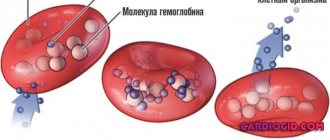
Myoglobin is a protein that contains iron (ferrous).
Myoglobin is identical in structure to hemoglobin, but their protein structure is different. This protein does not carry oxygen ions throughout the body, but stores it in the body, forms oxymyoglobin molecules, and fills muscle tissue with oxymyoglobin.
Muscle tissue cells filled with oxygen are able to ensure their internal respiration.
The oxygen consumption of muscle tissue depends entirely on its function.
Tissues that intensively consume oxygen:
- Cardiac muscle cells
- Gray matter, which is found in the brain,
- Parenchyma of the liver organ,
- The cortex, which is part of the kidneys.
Only the cells of one single tissue can accumulate oxygen molecules - muscle tissue, because it has myoglobin molecules in its composition.
What are muscle hemoglobin molecules?
Myoglobin is a protein that is present in striated muscles, similarly located in myocardial tissue. Myoglobin is part of a group of chromoprotein molecules.
Myoglobin contains a gene that is interconnected with part of the body’s proteins and is part of the prosthetic group. The myoglobin molecule contains amino acid components.
A protein is a monomer that consists of a single chain.
There are structures of a protein molecule:
- The primary structure is the monomer stage, consisting of a polypeptide linking chain, which includes amino acid residues,
- The structure of the secondary confirmation stage , which has an α-helical shape and 75.0% of molecules have this structure,
- The tertiary type structure has an α-helical shape, which is folded into a compact globule.
To determine the structure of a tertiary species, an X-ray analysis method is needed.
Properties of myoglobin
Myoglobin has unique abilities:
- Hemoglobin binding,
- Creating a custom buffer
- 1.0 grams of myoglobin can add 1.34 ml of o2,
- The ability to store oxygen reserves until the near future.
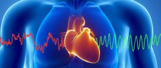
If a situation occurs in the body and the myocardium does not receive oxygen, then this supply of oxygen in myoglobin provides the heart muscle for 4 seconds.
When disruptions occur in the blood flow of the myocardium, or damage occurs in systole, then hemoprotein will prevent oxidation of the body and provide normal conditions for all processes in the body.
The work of myoglobin in this provision is short-term.
When skeletal muscle myocytes, or cardiomyocytes, are damaged, myoglobin is released into the bloodstream.
During myocardial infarction, the level of myoglobin in the blood depends on the degree of damage to the heart muscle by necrosis. The more the heart muscle is affected, the higher the muscle chromoprotein index in the blood.
When a myoglobin test is repeated during a heart attack (after 2-3 hours), then using this analysis the degree of destruction of the heart muscle can be determined.
Also, an increase in the level of creatine kinase (CF fraction) may indicate a heart attack.
Myoglobin in myocardial infarction
Although the content of aspartate aminotransferase
(AST) in the heart and the largest of all internal organs, this enzyme is also found in the brain, lungs, skeletal muscles, kidneys, liver and other organs and tissues. Therefore, an increase in AST activity in the blood is a sensitive but not specific marker of myocardial damage. Most laboratories refuse to determine it due to the availability and informativeness of the CPK determination, not to mention those cases where it is possible to determine cardiac-specific troponins.
Activity in the blood
Lactate dehydrogenase (LDH) increases more slowly during myocardial infarction and remains elevated longer than CPK or its MB fraction. This is a useful test for the retrospective diagnosis of myocardial infarction, when the patient is admitted to the hospital a day to a week after the onset of a coronary accident. True, many laboratories are increasingly using the determination of cardiac-specific troponins for this purpose.
Increase in total LDH
is not specific for myocardial damage. The total activity of LDH in the blood can increase in acute and chronic muscle pathology, pulmonary embolism, shock of any etiology, megaloblastic anemia, leukemia, liver and kidney pathology, as well as in a number of other diseases. Speaking about LDH isoenzymes, recall that LDH1 is found primarily in the heart and kidneys, while LDH4 and LDH5 are found in the liver and skeletal muscles. With hemolysis, overestimated values of LDHR can be obtained since this isoenzyme is also contained in erythrocytes.
Myoglobin
- an early marker of myocardial damage - appears in the blood plasma in the first hours after the development of myocardial infarction. However, interpretation of the results of myoglobin determination is difficult due to the nonspecificity of this marker (it is found in skeletal muscle). In other words, with an uninformative ECG, it is impossible to diagnose myocardial infarction based only on an increase in the level of myoglobin in the blood. This result should be “reinforced” by similar shifts in the concentration of the CF fraction of CPK or cardiac-specific troponins.
Read also: Myocardial infarction conclusion
Troponins
are regulatory proteins for muscle contraction. There are three types of them in the heart: C (“si”), I (“ai”), T (“ti”). Troponin C, which is found not only in cardiomyocytes, but also in smooth muscle fibers, is not suitable for diagnosing myocardial damage. For this purpose, the determination of troponin I or T in the blood is used. Although a small amount of the latter may be present in skeletal muscle, it is believed that currently used tests do not detect it in the blood.
Determination of troponins
increases the sensitivity of diagnosing myocardial damage. Figuratively speaking, this test allows you to determine the death of “counted” cardiomyocytes. Clinically, this is both good and bad. It’s good, because it allows us to confirm the development of even the smallest myocardial infarction or to identify a subgroup with an unfavorable prognosis among patients with unstable angina. In some of the latter, the duration of temporary obstruction of the coronary artery is sufficient for the development of cardiomyocyte necrosis without changes in the ECG and/or elevation of CPK characteristic of a heart attack. Determination of cardiac-specific troponins in patients with unstable angina serves as a measure of plaque instability.
It's bad because patients
with heart failure and/or myocardial hypertrophy against the background of arterial hypertension, the level of cardiac-specific troponins in the blood can also increase without the development of myocardial infarction. This, naturally, complicates the diagnosis of myocardial infarction in this group of patients. Let us note once again that an increase in the level of blood troponins is evidence of damage to cardiomyocytes of any origin (toxic, inflammatory, electrical - during cardioversion, thermal - during ablation) and not necessarily ischemic. The dynamics of the rise in troponins in blood plasma during myocardial infarction is similar to that of the CF fraction of CPK.
Dynamics of serum enzymes after a typical myocardial infarction. CPK - creatine phosphokinase; LDH - lactate dehydrogenase; GOT—glutamate xaloacetate transaminase.
What is the difference between this protein and hemoglobin?
Both proteins in the body, hemoglobin and the muscle protein myoglobin, have quite a few common components. Their main similarity is the transportation of oxygen ions throughout the body.
The function of myoglobin is to attach oxygen ions to itself and create oxygen reserves in tissues. Another function of myoglobin is to release oxygen from tissue cells to prepare energy for muscle function.
The structural components of these two myoglobin proteins and hemoglobin molecules are different.
Hemoglobin has a polymer structure, while myoglobin has a monomer molecule structure.
Differences:
Hemoglobin in the body is responsible for the respiration function of cells and maintains a constant pH level of blood acidity. In its structure it contains 4 molecular hemes.
The properties of myoglobin are the transport of oxygen ions from the lungs to the arteries of the peripheral sphere of the body. This protein has one chain in its structure, so it does not spend much effort to bind with oxygen molecules, although it is harder to give these molecules to it.
Both of these proteins are highly toxic when ingested in a free, pure state. Normally, myoglobin molecules are removed from the body by the kidneys.
Myoglobin molecules are large in size, so if there are disturbances in the vessels of the kidneys, they can clog them, causing necrosis of the organ.
Competition for oxygen occurs between these two proteins, and myoglobin does not have time to give it to tissue cells, which can provoke oxygen starvation of muscle tissue.
In cardiology, analysis of myoglobin is one of the brightest markers of the pathology of the cardiac organ, namely myocardial infarction.
This marker confirms a positive result of damage to the heart muscle, or muscle tissue of the body.
Myoglobin and methods for its determination in blood
Myoglobin is a special protein formation that is found in muscle tissue, including it is localized in the heart muscle.
It, like hemoglobin, has heme, which binds to a chain of amino acids. This substance belongs to the group of chromaproteins and is monomeric in structure, since it consists of only one chain.
What is this substance?
Currently, the following myoglobin structures have been studied:
- Primary - consists of one chain of polypeptides, which are amino acid residues.
- Secondary - in this form, ¾ of the chain has the form of an a-helical conformation;
- Tertiary - a secondary helix that folds into a globule.
The secondary and tertiary varieties of myoglobin were first discovered in the 60s of the last century. X-ray analysis was used to identify the latter species.
Myoglobin and hemoglobin have certain similarities in many respects. They are both proteins that take part in the processes of oxygen exchange by transferring it to tissue structures.
Myoglobin provides oxygen reserves in muscle tissue, and, if necessary, releases it when an acute need arises and for energy. All these processes create uninterrupted muscle function in various conditions.
Myoglobin and hemoglobin are somewhat different in structure . Myoglobin has a simpler structure, since it is a monomer, and hemoglobin is a polymer.
This protein, like hemoglobin, when it enters the blood in its free state, is very toxic. It enters the bloodstream as a result of injury with muscle rupture, or myocardial infarction.
In this case, the kidneys are responsible for the excretion of myoglobin, but since it is highly molecular, it causes blockage of the tubules and part of the renal parenchyma undergoes necrotic changes.
When myoglobin enters the blood, a process of competition between these two substances for free oxygen occurs, but since myoglobin releases it poorly, hypoxia develops.
This divergence of hemoglobin and myoglobin leads to the death of a person, as, for example, happens with tissue compression syndrome. Death occurs as a result of self-poisoning of the body.
Why do we need analysis and norm indicators?
Myoglobin testing is usually performed in the following cases:
- For clinicians, myoglobin readings in the blood are important evidence of the presence of myocardial infarction, since with the development of this disease, damage to the heart muscle and fiber necrosis occurs.
- Another indication is when the level of this protein substance is elevated due to compression or trauma. This may also be an indicator of a disorder in the functioning of the kidneys, or in other diseases that are accompanied by a violation of the integrity of the muscles.
The normal level of myoglobin in the blood in mcg/l is usually:
- from 19 to 92 in men;
- from 12 to 76 in women.
To determine this protein, there is an immunonephelometric analysis, immunofluorescence study or radioimmunological method. The final result is compared with the accepted norm.
If the sensitivity of the method for determining myoglobin is low, it may not be detected normally. It is not present in the urine of a healthy person.
When myoglobin is low or high
An increase in myoglobin in the blood occurs as a result of a violation of the integrity of muscle fibers in the following pathological processes and conditions:
- large area burns;
- traumatic injuries with muscle rupture;
- myocardial infarction;
- convulsive syndrome;
- renal failure.
A decrease in the level of this protein occurs in certain diseases:
- muscle weakness;
- rheumatoid arthritis;
- severe myositis;
- autoimmune pathologies.
When the level of myoglobin in the blood increases, it indicates the presence of myocardial infarction. And it is the main marker of this disease.
Within a couple of hours from the onset of chest pain, the analysis will show an increase in this indicator, and the highest numbers are determined after approximately 48 hours. This allows you to diagnose an acute condition and take all measures to relieve it in a timely manner.
The greater the area of damage to the heart muscle, the greater the quantitative results. A decrease in myoglobin after an acute attack is observed within 24 hours, provided that the process is suspended.
If the affected area continues to increase, then the level of this specific protein will also increase.
If there are two negative results in a row, the question of whether the patient has a heart attack is removed. In this case, you should look for another cause of chest pain.
Proper preparation for analysis
In order to detect myoglobin, you can take blood serum, plasma or urine for testing. All materials must be in freshly collected form.
In some cases, they are suitable for obtaining information if they are stored at a temperature of −25 degrees Celsius, but not more than two years.
To obtain true myoglobin numbers, the patient should follow some rules:
- the last meal should be 8 hours before the test;
- During this period, you can only drink clean water - juices, coffee, teas and other drinks are strictly prohibited;
- one hour before determining myoglobin in the blood, the patient is not allowed to smoke;
- for 30 minutes any physical activity is excluded;
- It is advisable to conduct the study against the background of an even psycho-emotional state;
- It is not recommended to do the analysis immediately after an ultrasound, x-ray examination, as well as after some physiotherapeutic treatments associated with electric pulse currents.
An analysis of blood myoglobin levels has important diagnostic value. Modern clinicians and cardiology specialists necessarily use it for differential diagnosis and confirmation of the effectiveness of treatment for myocardial infarction.
Sometimes this analysis is used in professional athletes. An increase in the level of this protein substance is evidence of severe muscle strain, and indicates the need to reduce the intensity of training.
When should a myoglobin test be done?
Blood biochemistry for the presence of myoglobin is done if it is suspected:
- Cardiac muscle infarction in the acute stage of pathology during its initial diagnostic study,
- Severe damage or destruction of skeletal muscle tissue,
- Pathology myopathy,
- The disease polymyositis,
- When monitoring the treatment of a heart attack.
In case of acute heart attack, blood for myoglobin is taken immediately upon admission of the patient to the clinic. Also to control the disease several times, at intervals of 3 hours.
The indications of this marker are very important in case of recurrent disease, and are prescribed in combination with other markers such as creatine kinase and troponin. This complex of biochemistry makes it possible to fully assess the degree of damage to the muscles of the cardiac organ.
Preparing for analysis
To study biological material for myoglobin, the following are suitable: blood serum, plasma, or even urine. All biological materials must be freshly collected.
In order to obtain the most correct result of myoglobin indicators, it is necessary to follow the rules before and during the collection of material:
- It is recommended to donate blood in the morning,
- Last meal at least 8-9 hours before venous blood sampling,
- Do not drink alcohol in the last 48 hours before submitting the material for analysis,
- You can only drink clean water before donation,
- Coffee, juices, tea, and other drinks are prohibited during this period to avoid distorted material,
- Smoking is prohibited at least 60 minutes before the test.
- Half an hour before the procedure, stop all activity,
- Before taking the test, do not be nervous and be in a calm state,
- Do not carry out the procedure immediately after x-rays, as well as after ultrasound,
- Do not test after physiotherapy.
Myoglobin analysis is an important diagnostic step, especially in the treatment of heart attack.
The results of the analysis make it possible to track the effectiveness of treatment, and if necessary, adjust the medication course.
Normal myoglobin index
Standard indicators may increase upward and depend on laboratory research methods:
- Diagnostic test – immuno-nephelometric,
- RIA radioimmunoassay analysis,
- Study using immunofluorescence analysis.
Despite the varying sensitivity of laboratory tests, the volume of myoglobin does not exceed the index from 65.0 to 80.0 μg/l.
Standard indicator:
- For representatives of the stronger sex from 19.0 to 92.0 μg/l,
- For the female body from 12.0 to 76.0 μg/l,
- The average normative coefficient is 49.0, and also more or less by 17.0 units (for males),
- The average standard for women is 35.0, and also more or less by 14.0,
- The concentration of myoglobin molecules in urine is less than the index of 20.0 μg/l; in an absolutely healthy body, myoglobin protein should not be present in urine.
When the myoglobin index in the blood differs from the normative units, then myoglobinemia is diagnosed.
If the presence of myoglobin in urine is detected during the analysis, then a diagnosis of myoglobinuria is made.
Index increased
Physiological increase in muscle type hemoglobin. The causes are related to the load on the skeletal muscle tissue, especially during sports and training, as well as when using physical therapy with electrical impulses.
A pathological increase in the index occurs in the following diseases:
- Damage to the heart muscle during myocardial infarction (hemoprotein increases, and the level of creatine kinase also increases). Myoglobin increases 30 minutes after the onset of pain, and its presence can be detected even 3 days after the attack,
- Kidney failure and uremic syndrome,
- Inflammation that occurs in muscle tissue
- In case of injury,
- Deep thermal tissue burns,
- Chemical burns in muscle tissue,
- Muscle cramps,
- After surgery,
- Muscle dystrophy.
Index downgraded
The muscle type hemoglobin index in the blood decreases only under the influence of pathology, such as:
- Arthritis of rheumatoid type,
- Inflammation of muscle tissue polymyositis,
- Myasthenia gravis is the presence of antibodies in the blood that affect myoglobin.
Polymyositis
Myoglobin in the blood - function and concentration norm
We will talk about what myoglobin is and what role it plays in the body. What blood concentration values are normal? What are the causes and risks of high or low protein levels in the blood and urine?
What is myoglobin
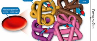
Myoglobin is a protein ball, i.e. a complex of proteins in which the constituent chains (peptides) are twisted on themselves in such a way that they form a helix with 8 sections. A structure that gives a molecule a spherical or spherical shape, hence the name globular protein.
Myoglobin's function is to transport oxygen to the mitochondria of muscle cells so they can contract.
How does myoglobin perform its function?
Myoglobin consists of a chain of 153 or 154 amino acids, which contain a heme group inside.
| The heme group is a chemical compound that contains an iron atom and is capable of binding to oxygen. From a chemical point of view, the heme group is a protoporphyrin, with the iron ion Fe2+ in the center. Protoporfinir is a compound of 4 pyrrole groups held together by 4 methylene bridges =CH−. |
The heme group, due to the presence of iron ion, allows myoglobin to stably retain an oxygen molecule. In addition, the special structure of the molecule results in the heme group and iron being enclosed in a kind of pocket, with hydrophobic properties and therefore able to stay away from water, which has oxidizing potential.
All this allows iron to maintain its oxidation state of 2+ and not switch to 3+. A condition that allows myoglobin not only to bind oxygen, but also to maintain a high degree of affinity for cells.
The main functions of myoglobin in the human body are:
- binds oxygen when its partial pressure exceeds 40 mmHg.
- releases oxygen when its partial pressure drops below 5 mmHg.
- the ability of myoglobin to bind oxygen regardless of temperature, pH, partial pressure of carbon dioxide (a function known as the Bohr effect).
Biological functions of myoglobin and difference from hemoglobin
From the above, one can already imagine myoglobin, an analogue of hemoglobin, which binds oxygen in the lungs and transfers it to cells to ensure metabolic processes.
Recall that cellular respiration is a long series of reactions in which nutrients (carbohydrates, proteins and fats) are oxidized to carbon dioxide, water and energy. The latter will be available to cells in the form of a chemical compound known as ATP:
C6H12O6 + 6O2 → 6CO2 + 6H2O + 38 ATP
There are of course important differences between these two globular proteins, both of which are capable of binding oxygen.
The differences that arise from differences in molecular structure can be summarized as follows:
- myoglobin has only one heme group (hemoglobin has 4) and can only hold one oxygen molecule versus 4 in hemoglobin.
- The ability to bind oxygen of these spherical proteins depends on the partial pressure: Hemoglobin binds oxygen at a pressure of about 90 mmHg and transmits it at a low pressure, but not lower than 40 mmHg.
- Myoglobin binds oxygen at pressures of the order of 50/40 mmHg and releases only at pressures of the order of 5 mmHg.
These differences lead to:
- Hemoglobin, contained in red blood cells, stores oxygen and transfers it to cells for metabolic processes in the body.
- Myoglobin, which is found mainly in muscle fibers, binds and stores oxygen, and then returns it to muscle tissue when the partial pressure of oxygen drops below 5 millimeters of mercury. Such conditions are achieved in the initial stage of muscle work and at the stage of intense effort, during which breathing cannot provide a sufficient flow of oxygen. In marine mammals, the concentration of myoglobin is so high that they can hold their breath for hours (whales).
Normal myoglobin levels in blood and urine
Myoglobin concentration can be measured in serum using a simple chemical test on a blood sample taken from a vein.
Normal myoglobin values vary from person to person and depend on muscle mass, age, and race.
| The maximum level of myoglobin in the blood is considered to be 90 nanograms per milliliter of blood, for both men and women. |
From the blood, myoglobin can be filtered by the kidneys and passes into the urine, where its concentration can also be measured.
| The maximum value of myoglobin in urine is considered normal at a level of 4 milligrams per liter of urine. |
Deviations in myoglobin levels
When the level of serum myoglobin differs from the norm, then they speak of myoglobinemia , and if a laboratory analysis reveals a high content of myoglobin in the urine, then they speak of myoglobinuria .
When the values are below the physiological norm, we speak of low myoglobin.
Causes of high myoglobin values in blood or urine
High myoglobin values are almost always observed after the breakdown of muscle fibers, resulting in its high availability in the blood. However, there are many reasons that can determine this condition, both physiological and pathological.
Causes…
The main ones are:
- Heart attack. In the presence of ischemia (complete or partial lack of blood flow) of the heart muscle, a rapid increase in myoglobin levels occurs. All this allows you to diagnose the likelihood of a heart attack by measuring myoglobin levels. Of course, this test is not specific.
Learn more about the causes and risk factors of myocardial infarction.
- Muscle injuries. All muscles can be in such a state that there is an increase in the level of myoglobin between muscle fibers.
- Excessive physical work, especially without the necessary training. For example, participating in a running marathon without preliminary training.
- Hereditary causes, that is, genetic factors.
- Burns with damage to large muscle masses.
- Kidney failure.
- Poisoning with chemicals and certain medications.
- Myositis, i.e. inflammation of skeletal striated muscles.
- Muscular dystrophy. A set of degenerative diseases that cause muscle atrophy due to a deficiency of the protein dystrophin, which is necessary for the formation of the physiological structure of muscle fibers.
| As blood myoglobin travels to the kidneys, where it is filtered and excreted in urine, the presence of high levels of myoglobin can lead to kidney failure because myoglobin is nephrotoxic and can damage the kidney tubules, which are part of the nephrons. |
Causes of Low Myoglobin Levels
Very low myoglobin values can be caused by:
- Immune system problems that can produce antibodies against myoglobin in the blood .
- Myasthenia. The disease is characterized by muscle weakness caused by a failure of the immune system, which produces antibodies that block cholinergic receptors, suppressing the production of acetylcholine, which is responsible for the transmission of nerve impulses.
However, this condition does not pose any particular health risk.
What affects muscle hemoglobin levels?
Several factors can influence the level of myoglobin in the blood, both upward and downward:
- Hemolysis,
- Lipemia affects the indicator of this protein in the analysis,
- Abuse of alcoholic beverages provokes an increase in myoglobin in the blood,
- Consumption of large amounts of amphetamines increases the level of this protein,
- Renal failure affects the level of myoglobin in the blood, since this substance is excreted from the body by the kidneys.
Myoglobin: 3 indications for analysis, norm, interpretation of the result
Myoglobin is a protein that contains iron, forming heme. The main location of this protein is muscle tissue. This type of tissue forms skeletal muscles, as well as the most important muscle in our body - the heart.
Muscle tissue forms the myocardium - the layer of the heart that ensures its contractile function.
The main task of myoglobin is to form a depot for oxygen. That is, if there is a lack of oxygen, myoglobin will supply it to the muscles, ensuring muscle contraction.
Difference from hemoglobin
Myoglobin is similar to hemoglobin. They are somewhat similar to each other, but there are also a number of differences.
https://www.youtube.com/watch?v=7Cf7ER-AL3E
Myoglobin and hemoglobin contain heme, which contains iron. It is this part of the molecule that allows it to bind to oxygen.
The difference between these proteins lies in the functions they perform. Thus, hemoglobin is a transport protein - it transports gases in the blood, including oxygen. And myoglobin stores this oxygen in reserve, which brought hemoglobin into the cells.
Collection of material for analysis
Determining the level of myoglobin in the blood is diagnostically significant. In this case, the material for research will be venous blood, from which serum will then be obtained.
It also makes sense to determine the presence of myoglobin in the urine.
The essence of the method
In modern laboratories, the determination of myoglobin in the blood is carried out using the immunoturbidimetric method. The essence of this method is to assess the degree of agglutination of myoglobin and special antibodies to it, which are connected to latex particles.
Latex particles allow visualization of the interaction between myoglobin and antibodies. This interaction changes the sample parameters, which are recorded on a special device. Then the level of myoglobin in the blood is calculated.
Due to the fact that myoglobin can enter the urine, it makes sense to determine it in this biological fluid. This can be a qualitative determination by adding ammonium sulfate. Ammonium sulfate will detect myoglobin in urine.
The detection of myoglobin in the urine is called myoglobinuria.
Dry chemistry methods, which are used in general urine analysis, can only determine the presence of blood in the urine. This may include any urine proteins that contain iron. Which is not the most informative method.
Electrophoresis is used to distinguish which specific proteins are found in the urine. Depending on their mass and charge, proteins will be distributed differently, making it possible to distinguish between them. This is important when diagnosing conditions that cause blood in the urine.
Indications for referral for analysis
In order to refer a patient to determine the level of myoglobin in the blood, there must be symptoms indicating a pathology of muscle tissue.
Myoglobin levels are determined when:
- suspected damage to the heart muscle as a result of a heart attack or other process;
- damage to skeletal muscles, for example, due to injury or inflammatory processes;
- assessing the severity of kidney damage.
Given the fact that myoglobin easily passes through the kidney filter, an excess of this protein in the blood can have a damaging effect on the kidneys.
Serum myoglobin levels differ between men and women. In children from the age of fourteen, the level of this protein becomes approximately the same as in adults.
In women, the normal concentration of myoglobin is 12–76 micrograms per liter (μg/L), and in men it is 19–92 μg/L. These data are indicative as they may vary in each laboratory depending on the reagent manufacturer.
Normally, myoglobin is almost undetectable in urine.
The level of myoglobin is so low that even if low concentrations are found in the blood, it has no diagnostic significance. Some laboratories consider the lower limit of normal to be 0 µg/L.
High concentrations
Increasing the concentration of myoglobin in the blood plays an important role in the diagnosis of a number of pathological conditions.
With myocardial infarction, there is an increase in myoglobin concentration within 2 hours. However, this indicator is not strictly specific for this pathology.
Creatine phosphokinase-MB and troponins are more specific markers of myocardial damage than myoglobin.
Also, if muscle tissue is damaged as a result of its death, injury, or impaired blood supply, an increase in the myoglobin content in the blood will be noted. This is observed in pathologies such as dermatomyositis, muscle dystrophy, etc.
During convulsions or after surgery, an increase in the concentration of myoglobin in the blood is observed.
Also, high concentrations of myoglobin in the blood lead to increased excretion in the urine, which will affect the level of this protein in the urine. Excess myoglobin will lead to kidney damage and the development of renal failure.
What can influence changes in myoglobin levels
There are a number of factors that can affect the level of myoglobin in the blood. These include:
- excessive physical activity;
- drinking alcohol on the eve of the test;
- donating blood on an empty stomach;
- intramuscular injections;
- taking Acetylcholine and Succinylcholine.
Therefore, before you go to donate blood, you should properly prepare and eliminate factors that could lead to an incorrect result.
Conclusion
Myoglobin is a muscle tissue protein that is involved in oxygen storage. When this tissue is damaged, the protein releases into the blood, which is reflected in blood and urine tests. Myoglobin helps in confirming the diagnosis of myocardial infarction, but is not the only indicator used for diagnosis.
Article rating
We have put a lot of effort into making sure you can read this article and would welcome your feedback in the form of a rating. The author will be pleased to see that you were interested in this material. Thank you!
(5 5,00 of 5) Loading...
If you liked the article, share it with your friends!
You might be interested
Presence of myoglobin in urine
The content of myoglobin in urine is an indicator of pathology in the body, since in a healthy person this protein is not visible in urine:
- Heart attack (damage by necrosis of the heart muscle),
- Myoglobinuria, as a secondary disease due to intoxication of the body,
- For large area burns and deep traumatic burns,
- Alcohol intoxication,
- Physical overload of muscle tissue is especially acute during sports activities,
- Toxicosis of muscle tissue due to trauma,
- Kidney pathology.
Conclusion
With proper distribution of the load on the muscle group and a healthy, balanced diet and lifestyle, myoglobin will be within standard values.
The role of this protein is very important in the human body: providing oxygen to muscle cells, transporting oxygen to its destination and transporting carbon dioxide to the lungs.
With a normal reading of myoglobin in the blood, there will be an uninterrupted operation of the gas exchange process in the body for the proper functioning of life-supporting organs.
(
1 ratings, average: 5.00 out of 5)
Interpretation of results
Norm:
Promotion:
- Myocardial infarction.
- Intense physical activity on the skeletal muscles (sports training).
- Conducting electropulse therapy.
- Skeletal muscle injuries – long-term compartment syndrome.
- Cramps.
- Inflammatory muscle diseases - myositis.
- Burns.
- Long course of intramuscular injections.
Decrease:
- Myasthenia gravis (the appearance of antibodies to myoglobin circulating in the blood).
- Polymyositis.
- Rheumatoid arthritis.
Select the symptoms that concern you and answer the questions. Find out how serious your problem is and whether you need to see a doctor.
Before using the information provided by medportal.org, please read the terms of the user agreement.