At certain intervals, people undergo a general blood test . The results of this clinical study show how well the body functions.
If any organs or systems fail, this almost always affects the quality of the blood. Today, patients present with microcytosis . Many people are frightened by such a diagnosis, without even understanding what it is. In fact, such a violation in itself is not dangerous. It only indicates that certain problems are occurring in the body.
What is macrocytosis, macrocytes
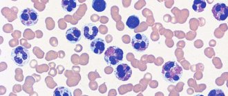
Macrocytes in a blood smear
Macrocytes are abnormally large cellular components of blood. The presence of such elements in the bloodstream is called macrocytosis. This condition is a consequence of a disruption in the process of hematopoiesis at the stage of cell generation. Incorrectly formed red blood cells are not able to perform their functions properly, that is, they cannot deliver hemoglobin through the capillaries, participate in the creation of immunity and metabolic processes.
In infants, macrocytosis is normal. And for pregnant women, it is a frequent companion throughout the entire period of bearing a baby, which is associated with the restructuring of processes in the bloodstream. Correcting nutrition and taking vitamins helps pregnant women cope with this disorder. For other groups of the population, this is a dangerous symptom.
In most cases, an unnatural increase in blood cells is accompanied by anemia - a low level of hemoglobin in the bloodstream. Anemia is a common symptom that reflects a number of diseases and is characterized by a lack of oxygen in the tissues of the body. An in-depth study of the causes and extent of macrocytosis allows us to determine the type of anemia and its treatment regimen.
Pathology in children
In the first months of life, an increased level of microcytes is observed, and this is normal. This is explained by the fact that the child’s organs are not fully formed.
This process is completed by about three months. If significant deviations are diagnosed, the child is hospitalized to exclude the development of congenital diseases.
In 1 year, the indicator should be completely normalized, otherwise a thorough examination is required. The primary degree of development indicates a violation of the immune system and requires adjustments in nutrition and the prescription of a vitamin complex.
In severe situations, indicates the development of leukemia.
Minor violations are normally allowed in adolescence, this is associated with hormonal levels and goes away on its own.
Macrocytosis in a blood test. Deviations in macrocytosis
Determining red blood cell parameters is an important diagnostic step
Comprehensive laboratory diagnostics allows you to determine the size of blood cells and their characteristics with high accuracy. To identify macrocytosis and the degree of its development, the following indicators are used.
- MCV—mean erythrocyte volume. It is measured in both femtoliters (fl) and micrometers (µm).
- RDW—red blood cell distribution width.
- MCH is the average hemoglobin content in a red blood cell.
- MCHC is the average concentration of hemoglobin in an erythrocyte.
- HGB is the hemoglobin content in the bloodstream.
- NBT is the ratio of red blood cells to its total volume.
Standard indicators of red blood cells:
- MCV = 6.2 - 8.2 µm;
- MCV = 80 – 94 fl;
- RDW = 11.5 - 14.5%;
- MCH = 27 - 35 pg;
- MCHC = 310 - 360 g/l;
- HGB = 120 - 140 g/l for women, 130 - 160 g/l for men;
- NCT = 0.36 - 0.46 l/l for women, 0.41 - 0.52 for men.
Based on the degree of deviation of the data obtained in the analysis, the stage of development of macrocytosis is determined. An additional study may include taking a bone marrow sample, which will help determine the cause of the pathology.
Analysis in pregnant women
Microcytosis is detected during a blood test. If the presence of anemia is determined, a smear of peripheral material is additionally prescribed.
Thanks to this measure, the initial symptoms of micro- and macrocytosis (the diameter of red blood cells exceeds the norm) or a mixed type of anisocytosis, combining signs of the first and second types, are revealed. Normo-, hyper- or hypochromia are detected in the same way.
A hematologist deals with the disease; it is he who prescribes further diagnostics, determines the cause, and the necessary treatment.
As additional diagnostic measures, tests are prescribed to determine celiac disease, blood and stool tests for the presence of the microorganism Helicobacter pylori.
The specialist clarifies in detail all the signs and symptoms present in the patient; it often becomes necessary to consult a gastroenterologist (if pain appears in the abdominal area). Sometimes the following diagnostic methods are needed:
- Transabdominal ultrasound examination.
- Endoscopy of the stomach and intestines.
- Computed tomography of the abdomen.
If adult women experience heavy periods with pain, it is necessary to exclude the presence of uterine fibrosis and other factors that cause heavy bleeding.
The term “macrocytosis” refers to a phenomenon in which large quantities of erythrocytes (red blood cells) are present in the blood, slightly or significantly changing their size towards an increase. Anemic syndrome, caused by the presence of a large number of enlarged formed elements of the erythrocyte series, is classified as macrocytic anemia.
Cells with a diameter of 8 microns and above fall into the category of macrocytes (the diameter of healthy red blood cells is ≈ 7-8 microns - normocytes, everything below is microcytes).
Meanwhile, in certain pathological conditions (lack of vitamin B12 and folic acid, for example), cells appear in the blood that are larger in size than macrocytes; their diameter is more than 10 microns - these are megalocytes. The anemia that results from this will also be macrocytic, but more often it is called megaloblastic in order to emphasize its origin.
Enlarged cells usually carry a larger amount of red blood pigment, chromoprotein - hemoglobin, so their color may be more intense than those red blood cells that are saturated with hemoglobin correctly or even less than necessary. This intense staining of macrocytes is called hyperchromia. Macrocytosis with hyperchromia are the main laboratory signs of hyperchomic macrocytic anemia.
blood is normal and with hyperchromic macrocytic anemia
The key point in the diagnostic search (in relation to macrocytic anemia) is a general blood test (hemogram), or rather, its individual parameters:
- Morphological characteristics of red blood cells (detection of macrocytes, poikilocytosis);
- MCV indicator (mean blood cell volume) - it shows values above 120 femtoliters with macrocytosis of erythrocytes;
- Hematocrit level (in severe vitamin deficiency anemia for a long time, the hematocrit drops to 30-25%, although in other forms of macrocytic anemia it may remain within normal values).
It should be noted that the nature of the process is significant, since it is the chronic course of the pathology that creates the preconditions for the disappearance of normal red cells and their replacement by macrocytes.
In patients suffering from anemic syndrome for a shorter period of time, the indicator may only slightly exceed the upper limit of normal values or even be within the normal range. Then you will have to take into account other circumstances that could create conditions for the development of macrocytic anemia. Macrocytosis, caused by the use of chemotherapy drugs or damage to the hepatic parenchyma, is usually mild or moderate.
blood for B12 deficiency anemia
- Poikilocytosis of erythrocytes;
- The presence of fragments of destroyed cells in the smear (schizocytosis);
- Hyperchromia;
- Changes in the “white” blood - a significant increase in the content of neutrophilic leukocytes, in the nucleus of which there are 5 or 6 segments, while normally the number of cells containing more than five segments in their nucleus is unlikely to reach 5% of the total number of segmented neutrophils .
The presence of a huge number of polysegmented neutrophil leukocytes is a kind of equivalent of poikilocytosis of red blood cells and is an important laboratory symptom of megaloblastic anemia.
With the onset of pregnancy, a number of changes occur in the body of the expectant mother. Joyful anticipation can be overshadowed by the deterioration of the general condition of the body, which often occurs against the background of anemia.
An alarming signal for doctors is a shift of red blood cells in the blood picture towards macrocytes. This happens primarily due to a lack of folic acid in the female body.
The main reasons leading to folate deficiency during pregnancy, experts include:
- an increase in blood mass (while the increase in plasma does not correspond to a quantitative increase in red blood cells);
- poor nutrition, leading to a decrease in the body’s absorption of B vitamins;
- multiple pregnancy;
- taking anticonvulsants;
- toxicosis of pregnant women.
A deficiency of folic acid in a pregnant woman's body can lead to placental abruption, premature birth and the birth of a low-birth-weight baby.
Anisocytosis of erythrocytes is a pathology that leads to a variety of disorders associated with changes in the content of various types of red cells. In almost all cases, this leads to a number of diseases that require additional diagnostics and therapy.
Red blood cells are responsible for transporting oxygen and carbon dioxide, as well as nutrients in the body.
This analysis is carried out using analyzers. They help to accurately count red blood cells of all sizes and shapes in 1 μl of blood. The sampling is carried out on an empty stomach. The material for the study is taken from a vein.
The average capacity of an erythrocyte is determined automatically, and the deviation in the number of normocytes in relation to macro- and microcytes is calculated. The data is displayed in the form of a histogram. If the percentage of RDW falls within the normal range, then the result is recorded as negative.
If the norm is exceeded, a positive result is indicated. For such cases, the procedure is duplicated in order to identify the causes of abnormal indicators.
Sometimes a false positive result occurs, especially if there are a large number of macrocytes in the blood. These results often occur after surgery or blood transfusion. The redistribution or change in the type of cells itself can change frequently, and in a short time.
Therefore, it may be necessary to process the data using a statistical analysis method. The standard deviation value is calculated using a number of formulas. Previously, the results were obtained manually, but this is an extremely labor-intensive process, and therefore today it has been practically abandoned in favor of computer data analysis.
The indicator of hypochromia is determined initially by a general blood test. To identify pathology, the doctor very carefully evaluates the color characteristics of the red blood cell. Normally it should be completely red. Color standard - 0.85 - 1.05.
When hemoglobin in the blood decreases, this is immediately indicated by the color of the red body - red with a white center. Such a cell visually resembles a target. This is hypochromia.
It can occur for various reasons: a lack of foods containing iron in the diet, or it can occur due to frequent but slight blood loss.
For this pathology, special therapy is prescribed, which involves adjusting the diet and taking special iron-containing drugs.
The symptoms of macrocytic anemia are not clearly expressed, so the disease is often diagnosed only after certain diagnostics.
With megaloblastic macrocytosis, red blood cells change degeneratively, that is, the shape of the cells may be disrupted (poikilocytes) or the structure may be destroyed (schizocytes).
Symptoms of macrocytosis
Visual impairment is a possible sign of macrocytosis
At an early stage, symptoms may not appear. Changes in the size of blood cells are often discovered by chance during a routine blood test. Later stages are characterized by the following symptoms:
- fatigue, weakness;
- sudden weight loss;
- varnish tongue (with a smooth, shiny surface);
- pallor or yellowness of the skin;
- fainting;
- dyspnea;
- cardiopalmus;
- tingling in the limbs;
- brittleness of hair and nails;
- flashing “flies” before the eyes;
- dizziness.
All these symptoms are characteristic of the state of anemia, and each symptom separately helps to determine what exactly the body lacks at the moment to restore oxygen metabolism. Brittle nails and hair indicate a lack of iron, a varnished tongue indicates a lack of vitamin B 12.
Symptoms of macrocytosis appear cyclically, that is, the patient notices them periodically, and not constantly. This often leads to the fact that a person simply gets used to it and does not pay attention to changes in his body. Meanwhile, any of these symptoms is a reason to consult a doctor.
Hypochromic microcytic anemia in a child
In the zone of special control are the hemogram indicators belonging to the growing organism. Hypochromia and microcytosis in a general blood test plus other signs of trouble (excessive weight gain or loss, an unnatural need to taste and even eat inedible foods, changes in behavior, decreased concentration) make one suspect that the child is developing an anemic condition due to insufficient content in the body iron, which is so necessary for the synthesis of the red blood pigment - hemoglobin (Hb). And a decrease in the content of hemoglobin, the carrier of oxygen throughout organs and tissues, will entail the undesirable consequences that constitute the symptoms of IDA.
It is known that such disorders in children's bodies occur much more often than in adults, whose life support systems have already completed their formation. The thing is that this situation arises due to the peculiarities of iron metabolism and nutrition in children. For example, in a child who has just been born, the level of this chemical element (Fe) is 10 times lower than its content in the body of an adult, so for the first 15 years there is a constant replacement of the deficiency, which is ensured by absorption in the gastrointestinal tract from 0.8 to 1, 5 grams daily. And here the main hope is in the diet, because it is she who must “take care” that there is enough iron for the synthesis of hemoglobin.
A child under one year of age has the best chance of getting a normal amount of the element by drinking mother's milk, from which Fe is absorbed much more efficiently than from cow's or goat's milk.
In the future (after a year), the baby’s diet is also not particularly rich in iron, so to prevent IDA, it is advisable to pay attention to special foods from which the child’s body can take the amount of the element it needs. Otherwise (under conditions of instability in relation to iron that is natural for this age), microcytic anemia (IDA) will not take long to appear. If there are clinical signs of an anemic condition, IDA will be clearly “read” from the blood picture, first of all, making itself known by a decrease in hemoglobin levels.
Causes of macrocytosis
Pathology of the thyroid gland can cause macrocytosis
A detailed examination of the body allows us to determine the cause of the pathology.
The main reasons are:
- Lack of cyanocobalamin (vitamin B 12),
- lack of folic acid (vitamin B 9),
- dysfunction of the thyroid gland,
- hepatitis,
- intestinal diseases,
- damage to liver tissue,
- bone marrow pathologies,
- spleen diseases,
- some forms of leukemia,
- use of medications (Chloridine, Allopurinol).
Macrocytosis can also be caused by congenital anomalies. But often it is poor nutrition or alcohol abuse that becomes hostage to disorders.
Start by eliminating the cause
Macrocytosis of erythrocytes itself, of course, is not treated, because it is just a symptom identified in a general blood test. Treatment begins with eliminating the cause that created the formation of large cells. Meanwhile, it is not always possible to quickly eliminate the culprits of the misfortunes that have befallen them. Then long-term therapy begins for the doctor and the patient, aimed at the underlying pathology that caused the development of macrocytic anemia.
With vitamin deficiency conditions, everything is more or less clear; they are treated with appropriate medications (medicinal forms of cyanocobalamin and folic acid). However, if acquired deficiency (lack of food, malabsorption in the intestines, the influence of alcohol, competitive absorption by helminths, etc.) is easily amenable to therapeutic influence, then congenital anomalies react poorly to treatment, moreover, they are often accompanied by mental retardation and other abnormalities (megaloblastic anemia in children). Here we have our own tactics.
As for the entire list of diseases that gave rise to the formation of abnormally enlarged red blood cells in the bone marrow or to their premature and intense breakdown in the blood, in each case there is a separate approach: in some cases blood transfusions (blood transfusions) will help, in others they will be required surgical intervention.
Unfortunately, it is not only quite difficult, but also impossible, to describe all the options for the treatment process, however, readers can find answers to their questions regarding certain types of macrocytic anemia in the relevant publications posted on the pages of our website.
Treatment of macrocytosis
The cause of macrocytosis must be identified
Abnormally large blood cells are an indicator of the presence of the disease. Therefore, it is necessary to treat the root cause, and not the consequence of the development of the disease. The basis for restoring natural processes in the bloodstream is good nutrition, as well as giving up alcohol and smoking. A diet poor in vitamins provokes disturbances in blood formation, and alcohol-containing drinks lead to impaired liver function. Cigarettes contain poisons that contribute to intoxication of the entire body.
Additionally, depending on the disease, the following may be prescribed:
- course of vitamins,
- immunoglobulins,
- iron-containing preparations,
- blood transfusion,
- therapy with glucocorticoid drugs.
Severe forms of disease require mandatory hospitalization of the patient.
Prognosis and possible complications
Macrocytosis in childhood is also unsafe
Macrocytosis is a dangerous sign of a number of diseases characterized by a lack of oxygen in the body.
Immune suppression is one of the first consequences of this disorder, which leads to frequent infectious diseases and their complications.
In order to speed up the delivery of hemoglobin to the organs, the heart muscle begins to contract more often. The result is heart failure and tissue wear.
One of the common complications is a disorder of the central nervous system, which is manifested by impaired concentration and memory.
Lack of oxygen also leads to deformation of the structures of the mucous membranes and skin.
In childhood, the impact is on mental and physical development.
A condition in which tissues are not fully saturated with oxygen over a long period gradually leads to their degeneration. Severe forms of the disease lead to heart attacks, cardiogenic shock, and acute renal failure.
Timely treatment and dietary correction allow you to restore the natural circulation of red blood cells. However, in the future you should regularly monitor your health and undergo examinations.
Prevention
A balanced diet is a good way to prevent macrocytosis
Preventive measures are aimed at optimizing nutrition and giving up bad habits.
- Food must contain a sufficient amount of vitamins. However, you should not prescribe vitamin therapy yourself. An excess of vitamins and their incorrect combination lead to negative consequences, including macrocytosis.
- Smoking, in addition to intoxicating the body, poses a risk of developing macrocytosis due to the fact that cigarette smoke contains carbon monoxide. This component instantly combines with hemoglobin. As a result, hemoglobin molecules are no longer able to deliver oxygen to the tissues.
- Alcohol interferes with the absorption of microelements, including iron and vitamins, and also destroys hemoglobin molecules. Alcohol has a particularly negative effect on the liver, which leads to the accumulation of toxic substances in it and subsequent disruption of the kidneys, heart and brain.
- Active pastime in the fresh air will bring great benefits. This is the most effective way to saturate the body with oxygen.
Macrocytosis is not uncommon and is treatable in most cases. A healthy lifestyle and nutritious nutrition is a chance to forever get rid of the risk of a recurrence of pathology.